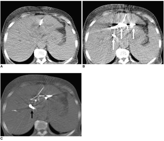
Fig. 2.
72-year-old patient with CBD cancer.
A. Using CT fluoroscopy image, left IHD is successfully punctured (bile runs out from needle hub).
B. Next, contrast media is injected to confirm if needle tip is located in bile duct (white arrows).
C. In spite of window setting control, exact identification of guide wire tip was impossible because of highly attenuated contrast media in hilar duct and CBD (arrow).

