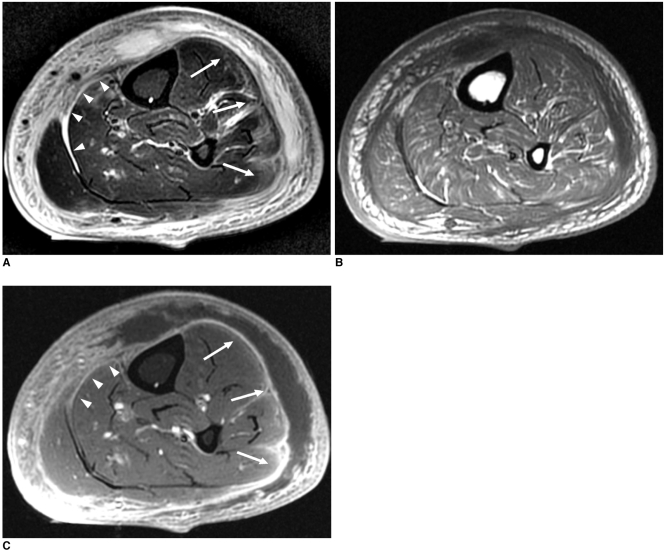
Fig. 1.
51-year-old man with surgically confirmed necrotizing fasciitis.
A. Axial fat-suppressed T2-weighted image (TR/TE, 4,000/87) shows peripheral band-like high signal intensity (arrows) in involved muscles of anterior, lateral and posterior compartments of left lower leg. Hyperintense signal is seen along deep fascia (arrowheads) as well as along superficial fascia.
B, C. Axial T1-weighted image (TR/TE, 450/12) (B) and fat-suppressed contrast-enhanced T1-weighted image (550/12) (C) show peripheral band-like enhancement (arrows) of involved muscles. Thin smooth enhancement is seen along deep fascia (arrowheads) as well as along superficial fascia. Patient underwent emergency fasciectomy. Streptococcus pyogenes was cultured.

