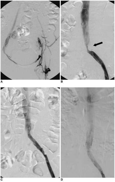
Fig. 2.
Treatment of iliac vein compression syndrome with deep vein thrombosis.
A. Venography showing thrombosis (fresh thrombus and 8 days after onset) and occlusion of left iliofemoral vein as well as contralateral venous drainage via pelvic venous collaterals.
B. Venography after thrombectomy and stenting showing patent left femoral vein and in-stent stenosis due to iliac vein compression (black arrow).
C. Venography after intra-stent percutaneous transluminal angioplasty showing widely patent left common iliac vein.
D. Venography one year after retrieval of filter showing remaining patent inferior vena cava and left iliofemoral vein.

