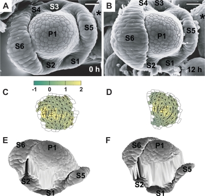
Fig. 11.
Flower primordium in which six sepals have been formed. Scanning electron micrographs (A, B), curvature plots (C, D), and side views of the reconstructed surface (E, F) show the portion of periphery of the clv3-2 inflorescence shoot apex No. 2, different from the portion shown in Fig. 4. Sepal primordia (S1–6) and the meristem (*) are labelled. The cellular pattern on the primordium periphery in the reconstruction (F) is missing due to the strong steepness of this primordium portion. Bars=20 μm. (This figure is available in colour at JXB online.)

