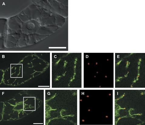
Fig. 4.
Transiently expressed RFP–ARP3 decorates sites of actin nucleation during recovery from cold-induced elimination of actin filaments in tobacco BY-2 cells that stably express GFP–FABD2. (A) Differential interference contrast of the target cell. (B–E) Co-localization of RFP–ARP3 and GFP–FABD2 in a focal section of the cell cortex. (F–I) Co-localization of RFP–ARP3 and GFP–FABD2 in a focal section of the cell centre. Details boxed in B and F are shown in C and G for the GFP–FABD2 fluorescence, in D and H for the RFP–ARP3 fluorescence, and in E and I for dual fluorescence, respectively. Bars=20 μm.

