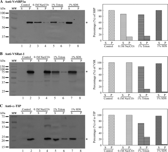
Fig. 9.
VrSBP1 is membrane-associated. The P100 vesicle fraction from developing mung bean cotyledons were first resuspended in solutions containing one of the three chemicals (0.1 M Na2CO3, 1% Triton X-100, and 1% SDS), as indicated, and incubated for 1 h at 4 °C, followed by centrifugation at 100 000 g for 1 h, resulting in pellet (P) and supernatant (S). The pellet (P) was resolubilized in the same volume of extraction solution containing 1.5% SDS and 150 mM NaCl. Equal volumes of protein samples were separated by SDS-PAGE, followed by Western blot analysis using anti-VrSBPa (A), anti-VSRat-1 (B), and anti-α-TIP (C), as indicated. Asterisks indicate the position of the detected proteins. The signals of the detected protein bands were further quantified with the software Quantity One (Bio-Rad) to determine the percentages of protein distribution between the pellet (P) and supernatant (S) in each treatment. MW, molecular weight marker in kDa.

