Figure 7.
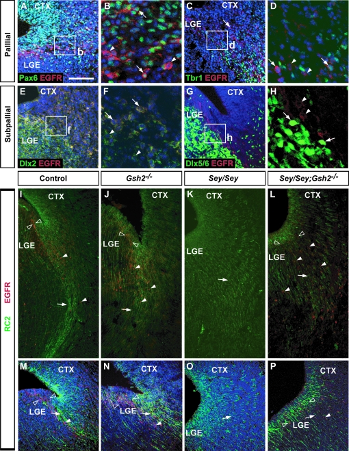
Alterations in LCS cell migration in Gsh2, Sey, and Sey/Gsh2 double mutants at E15.5. Low magnification of dual immunolabeling for Pax6 (green, A), Tbr1 (green, C), Dlx2 (green, E) and EGFR (red) shows coexpression of a subset of cells at the PSB (A, C, E). Higher magnification of boxed regions in (A, C, E) show individual double EGFR+ cells (arrows) colocalized with Pax6 (B, arrows), Tbr1 (D, arrows), or Dlx2 (F, arrows) intermingled with EGFR+ cells not expressing any of these markers (arrowheads). In contrast, there is no colocalization of endogenous Dlx5/6-GFP labeling (green, arrows) and EGFR immunolabeling (red, arrowheads) (G, H). RC2 (green) and EGFR (red) dual immunolabeling reveals the LCS radial glial scaffold (arrows) and EGFR+ migratory cells (arrowheads) emanating from the PSB (empty arrowheads) (I, M). In Gsh2 mutants, the VZ EGFR+ domain appears expanded (empty arrowheads), with a more diffuse RC2 immunolabeling (arrows) (J, N). EGFR+ cells (arrowheads), although disorganized, remain present along the LCS. In Sey mutants, EGFR+ cells are completely absent, and the RC2+ radial glia along the LCS migratory route is missing (K, O, arrows). In Sey/Gsh2 double mutants, RC2+ radial glia (arrows) resemble Sey mutants, but EGFR+ cells are now present along the LCS (arrowheads) and occupy an expanded domain at the PSB (empty arrowheads) (L, P). To-Pro-3 counterstaining (blue) is shown in the majority of panels. n numbers are as follows: EGFR and Pax6 or Dlx2 or Tbr1, n= 2; EGFR/Dlx5/6-GFP, n= 2; EGFR/RC2: controls, n= 3; Gsh2−/−, n= 3; Sey/Sey, n= 4; Sey/Sey;Gsh2−/−, n= 3. Scale bar: A, C, E, G, M–P: 100 μm, I–L: 40 μm, B, D, F, H: 30 μm.

