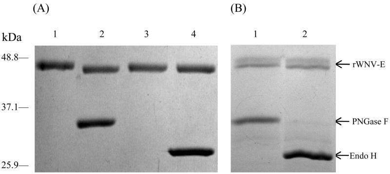
Fig. 2.
rWNV-E glycosylation pattern assessed in glycosylation assays that utilized peptide N-glycosidase F (PNGase F) or Endoglycosidase H (Endo H). On a Coomassie blue-stained reducing SDS-PAGE: (A) Drosophila-cell produced rWNV-E digested with either PNGase F (lane 2) or Endo H (lane 4). Lane 1 and 3 correspond to non-digested controls. Only PNGase F induced a slight gel shift of ~ 1kDa (lane 2), confirming N-linked glycosylation. (B) Baculovirus-produced rWNV-E was separately digested and then mixed with non-digested controls with either PNGase F (lane 1) or Endo H (lane 2). Both PNGase F and Endo H induced a gel shift > 3kDa. rWNV-E (arrow) upper and lower bands respectively correspond to the non-digested and the digested protein forms.

