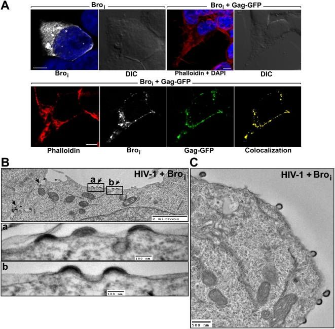
Figure 5. Broi is recruited by Gag to the plasma membrane and interferes with HIV-1 budding.
(A) First two upper left panels show a cell expressing HA-Broi (white). The next panel shows a reconstructed 3D view of a stack of Z sections from a cell expressing HA-Broi and Gag-GFP. Lower panels show a single Z section of the same cell showing colocalization of Broi and Gag-GFP at the plasma membrane. Broi (white) was stained with a mouse monoclonal anti-HA antibody and an Alexa 633-conjugated anti-mouse antibody. Nuclei were counterstained with DAPI (blue). F-actin was stained with Alexa 568-conjugated phalloidin (red) to delineate cells. The colocalization channel (yellow) of Broi and Gag-GFP was built using Imaris software. Scale bar = 5 µm. (B, C) Electron micrographs of 293T cells co-transfected with pNL4-3 wt and HA-Broi. (B) Arrested budding structures are indicated with black arrows. Two regions of interest in (a) and (b) show budding structures carrying electron-dense crescent-shaped material at a higher magnification. (C) HIV-1 budding structures tethered to the plasma membrane.

