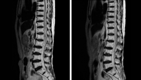
Fig. 1.
Sagittal T2-weighted magnetic resonance images showing high signal intensity within the spinal cord from the level of ninth thoracic vertebra to the conus medullaris that are indicative of diffuse spinal cord swelling, and fine signal voids around the cord surface that are indicative of vascular structures.

