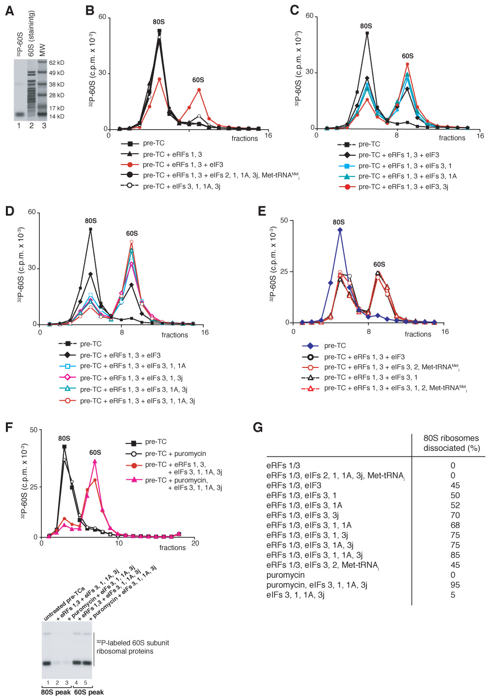
Figure 3. Dissociation of post-TCs into subunits.

(A) Coomassie staining of 60S subunit proteins (lane 2) and autoradiography of [32P]60S subunits phosphorylated by CKII (lane 1). MW markers are indicated. (B–F) Dissociation of pre-TCs, assembled on MVHL-STOP mRNA with [32P]60S subunits, after incubation with eRFs, eIFs and puromycin, as indicated, assayed after sucrose gradient centrifugation by Cerenkov counting and Pisarev, gel electrophoresis (F, right panel). The positions of 60S subunits and 80S ribosomes are indicated. (G) Summary of dissociation of post-termination ribosomes into subunits by different combination of eIFs (panels B–F).
