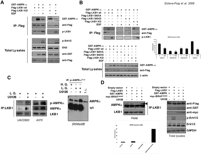
Figure 4. Growth factor treatment and BRAFV600E promotes LKB1-AMPKα disassembly.
(A) 293T cells were transiently transfected for 48 h with Flag-LKB1, Flag-LKB1KD (kinase dead) and GST-AMPKα as indicated. Then, cells were treated with 100 ng/ml of EGF for 10 min. Immunocomplexes pulled down with an anti-Flag-resin were separated by SDS-PAGE and proteins present in the complexes were analyzed by western blot. Total lysates show the transfection controls and the response to growth factor treatment. (B) 293T cells were transiently transfected with the constructs indicated. Then, cells were serum starved for 2 h and treated with 100 ng/ml of EGF for 10 min and protein complexes were immunoprecipitated with anti-Flag-resin. Protein complexes were separated by SDS-PAGE. Levels of GST-AMPKα, Flag-LKB1 constructs and the phosphorylation state of LKB1Ser431 in the complexes are shown. Quantification of the amount of GST-AMPKα normalized to the Flag-LKB1 immunoprecipitated is represented in the graph. Total lysates are shown for control transfection of the different samples. (C) Endogenous LKB1 from UACC903 and A375 melanoma cells growing in low glucose medium (L.G.) with or without 10 µM U0126 was immunoprecipitated. Western-blot from the immunoprecipitated samples was probed against LKB1, AMPKα and p-AMPKαT172 antibodies. On the right, total lysates from SKMel28 melanoma cells growing in complete medium (High Glucose, H.G.) low glucose medium (L.G.) in the presence or absence of 10 µM U0126 were subjected to immunoprecipitation with the anti-p-AMPKαT172. Samples were separated by SDS-PAGE. Total lysates (T.L.) from low glucose plus U0126 treated cells are showed as a control. Western-Blot of the immunoprecipitated samples was performed against total AMPKα antibody. (D) Hela cells were transfected with Flag-LKB1, GST-AMPKα and myc-BRAFV600E or and empty vector as indicated. Flag-LKB1 was immunoprecipitated and western-blots from immunoprecipitated samples were probed against the indicated antibodies. Graph shows the quantification of the AMPK bound to LKB1.

