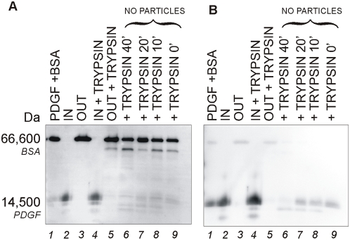
Figure 9. Immunoblot analysis showing that core-shell particles protect captured PDGF from tryptic degradation.
(A) Sypro ruby total protein staining and (B) Immunoblot analysis with anti-PDGF antibody of the same PVDF membrane are presented. Lane 1) control PDGF+BSA solution; 2) content of particles incubated with PDGF+BSA (IN); 3) supernatant of particles incubated with PDGF+BSA (OUT); 4) content of particles incubated with BSA+PDGF+trypsin (IN+TRYPSIN); 5) supernatant of particles incubated with BSA+PDGF+trypsin (OUT+TRYPSIN); 6) BSA+PDGF+trypsin without particles incubated for 40 minutes (+TRYPSIN 40′); 7)) BSA+PDGF+trypsin without particles incubated for 20 minutes (+TRYPSIN 20′); 8)) BSA+PDGF+trypsin without particles incubated for 10 minutes (+TRYPSIN 10′); 9)) BSA+PDGF+trypsin without particles incubated for 0 minutes (+TRYPSIN 0′).

