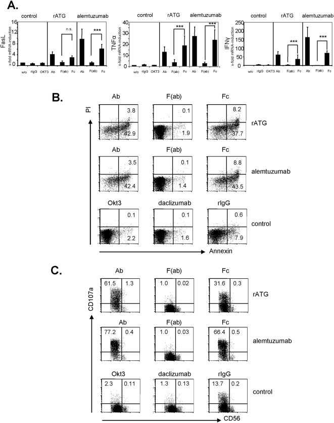
Figure 7. The IgG1 Fc-part of rATG and alemtuzumab is sufficient to induce enhanced cytokine expression, apoptosis and degranulation.
IL-2 (200 IU/ml) preactivated NK cells were incubated for 1 hour with either 1 µg/ml whole IgG antibodies, Fc-parts or F(ab) fragments of rATG or alemtuzumab. Cells treated with anti-CD3 (OKT3, IgG2a) or rabbit IgG served as controls. (A) Values for FasL, TNFα and IFNγ mRNA demonstrate the results relativized to untreated controls (2−ΔΔct) and are displayed as means±SD (n = 5): ***p>0.001. (B) Preactivated NK cells with IL-2 (200 IU/ml) were incubated with either 10 µg/ml intact antibody, Fc-parts or F(ab) fragments of rATG and alemtuzumab, or OKT3 for 1 hour. The antibodies daclizumab and rIgG served as controls. FACS dot plots illustrate staining for Annexin V and PI of treated CD56+CD3− NK cells. One representative of four independent experiments is shown. (C) Preactivated NK cells with IL-2 (200 IU/ml) were incubated with 10 µg/ml antibody preparations and anti-CD107a mAb for 3 hours. FACS dot plots illustrate staining for CD107a of CD56+CD3− cells. One representative of four independent experiments is shown.

