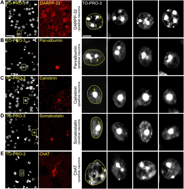
Figure 3. The various striatal neuronal populations have distinct nuclear morphology.
Mouse striatal slices were labeled with various markers specific for each type of striatal neuron and analyzed by confocal microscopy. For each row, the left panel shows a double staining with an antibody specific for a neuronal subtype and TO-PRO-3 (scale bar: 25 µm), the right panels are high magnification of TO-PRO-3-stained neurons (scale bar: 5 µm). For the first high magnification picture, which corresponds to the boxed area in the left panel, the dashed line follows the nuclear envelope and the number of clumps of highly compacted chromatin is indicated. (A) Neurons positive for DARPP-32, a marker of medium-sized spiny neurons, have a rounded nucleus about 10–11 µm of diameter with ≥3 clumps of intense TO-PRO-3 staining. (B–D) GABAergic interneurons. (B) Parvalbumin-positive interneurons have slightly elongated nuclei of 9–11 µm of diameter. Chromatin clumps are frequently observed surrounding the nucleolus, forming 1 or 2 clusters. (C) Nuclei of calretinin-positive interneurons are small (6–10 µm), irregular but mostly circular and have a unique central clump of heterochromatin. (D) Somatostatin-positive interneurons often have elongated nuclei (longest axis 8–12 µm, axis ratio ≥1.5) with a central clump of dense chromatin. (E) Cholinergic interneurons, identified by choline acetyltransferase (ChAT) immunoreactivity, have the largest nuclei (10–13 µm) with diffuse and irregular TO-PRO-3 staining with small clumps of intense fluorescence.

