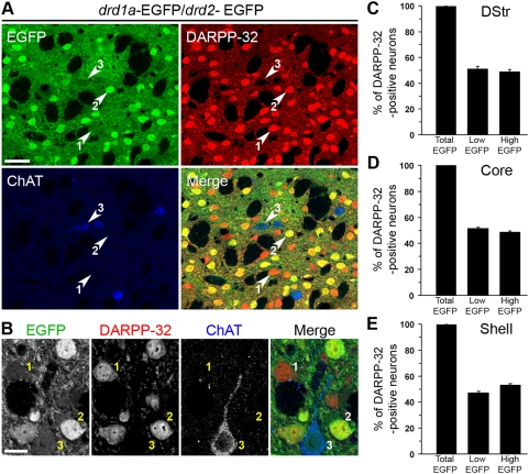
Figure 5. All MSNs express EGFP in drd1a-EGFP/drd2-EGFP double transgenic mice.
(A, B) Striatal slices from drd1a-EGFP/drd2-EGFP mice were studied by triple labeling for DARPP-32 immunoreactivity (red), ChAT immunoreactivity (blue) and EGFP autofluorescence (green). EGFP fluorescence was classified as high or low (see Fig. S2). Note that all high-EGFP neurons are MSNs, since they are always colabeled for DARPP-32, whereas low-EGFP neurons can be either MSNs (labeled for DARPP-32) or cholinergic interneurons (labeled for ChAT). Neurons could be therefore classified into three categories: 1, low EGFP/DARPP-32-positive (D1R-expressing MSNs); 2, high-EGFP/DARPP-32 positive (D2R-expressing MSNs, possibly coexpressing D1R); and 3, low EGFP/DARPP-32-negative (cholinergic interneurons expressing D2R). (B) Higher magnification showing neurons in the three categories defined above. Images are single confocal sections. Scale bars: 40 µm (A), 10 µm (B). (C–E) The percentage of DARPP-32-immunoreactive neurons showing any (Total EGFP), low (Low-EGFP) or high (High-EGFP) EGFP fluorescence in the dorsal striatum (DStr, C), nucleus accumbens core (Core, D) and shell (Shell, E). All EGFP neurons that do not show DARPP-32 immunoreactivity were identified as cholinergic interneurons. Data are means±SEM; n = 3 mice; 967 (C), 1708 (D) and 1271 (E) cells counted.

