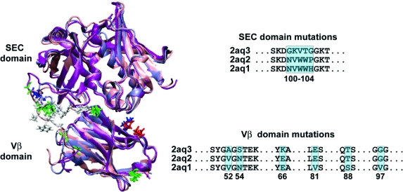
Figure 1.
The three simulated systems are structurally aligned for comparison. The SEC domain and Vb domain are shown in cartoon representation, with the mutated positions shown in licorice (hydrophobic residues in white, polar in green, negatively charged in red, positively charged in blue). An excerpt of the full sequence alignment is shown with mutated positions highlighted and numbered.

