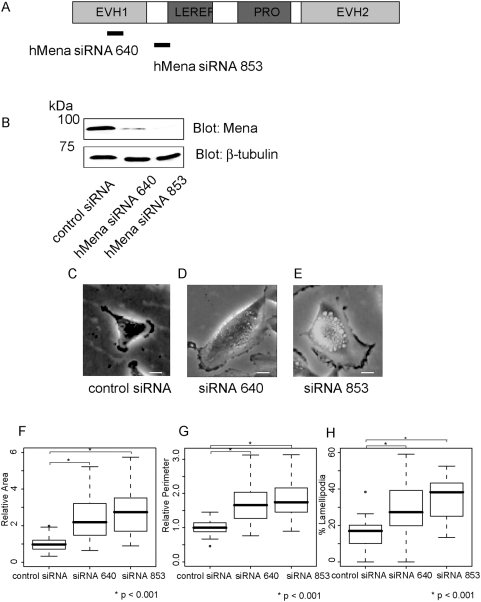
Figure 3. Reduced Mena expression induces lamellipodia formation and cell spreading in U251MG cells.
(A) Schematic of hMena. hMena contains a central proline-rich core flanked by three highly conserved regions, the EVH1, EVH2 and LERER domains. (B) Western blot analysis of U251MG cells shows reduced levels of the hMena protein in the cells transfected with hMena siRNAs (siRNA640 or siRNA853) compared with the cells transfected with the control siRNA. β-tubulin served as a loading control. (C–E) U251MG cells with siRNAs targeting hMena show spread, and increased formation of the lamellipodia. Bars, 10 µm. (F) Box and whisker plots for relative cell area of U251MG cells. The mean area of U251MG cells transfected with control siRNA was set as 1. (G) Box and whisker plots for the relative perimeter of the control cells and knock-down cells. The mean perimeter of U251MG cells transfected with control siRNA was set as 1. (H) Box and whisker plots for percent lamellipodial length of the perimeter. Data is from 50 cells each. For box and whisker plots, the top and bottom of the box represent the 75th and 25th quartile, and whiskers 10th and 90th percentiles, respectively. The middle line of the box is the median. Brackets with asterisks indicate statistically significant differences between the data sets from a Student's t test (p<0.001).

