Figure 4.
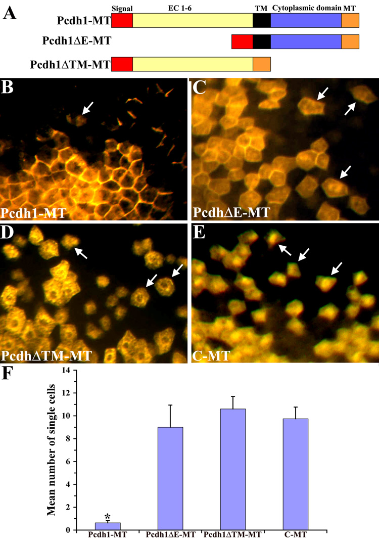
Ectopic expression of Pcdh1-MT promotes cell adhesion in a Xenopus cell mixing assay. A) Schematic diagram of the Pcdh1 constructs used in this study. The construct Pcdh1-MT encodes the full-length Pcdh1 protein. In Pcdh1ΔE-MT, the Pcdh1 extracellular domain (EC) is deleted and in Pcdh1ΔTM-MT, the transmembrane (TM) and cytoplasmic domains are deleted. All constructs are tagged with a C-terminal myc-epitope tag (MT). B–E) Xenopus embryos at the 16-cell stage were injected into a single dorsal blastomere with RNA encoding Pcdh1-MT (B), Pcdh1ΔE-MT (C), Pcdh1ΔTM-MT (D), or solely the myc-epitope (C-MT) as a control (E), then fixed at neurula stage and immunostained with the myc antibody. Arrows indicate single labeled cells not in contact with another labeled cell. F) The number of single labeled cells in the ectoderm was quantified for each construct. As compared to control embryos, embryos injected with RNA encoding Pcdh1-MT exhibited significantly fewer isolated cells, with the majority of Pcdh1-MT expressing cells remaining as a cohesive clone. In contrast, neither of the deletion constructs resulted in a decrease in the number of isolated cells, as compared to the control. * Indicates a statistically significant difference (p<0.01) from the control C-MT.

