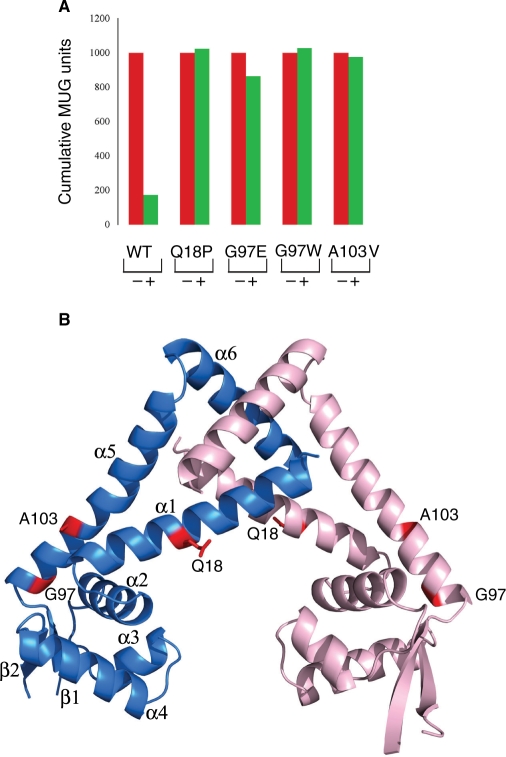
Figure 6.
Functional characterization of clinically isolated or in vitro selected MepR mutants. (A) Histograms of MUG units obtained from β-galactosidase assays for various MepR mutations. Measurements were made in the presence (+) and absence (–) of tetracycline, the addition of which causes MepR production. (B) Structural mapping of MepR mutations. The side chains of the mutated residues are shown as red sticks and labelled. One subunit is colored pink and the other blue. The secondary structure elements of one subunit are labelled.

