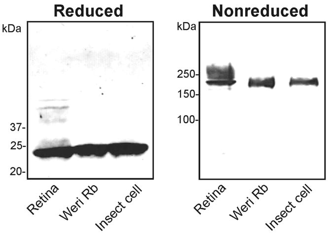
Figure 6.
Analysis of RS1 secreted from stably transformed Sf21 insect cells. The medium from Sf21 cells (insect cell) or Weri-retinoblastoma cells (Weri Rb) were immunoprecipitated on a RS1 3R10 antibody-Sepharose matrix. These samples together with RS1 from retinal extracts were separated on reducing and nonreducing SDS gels. Similar amounts of RS1 were applied to each lane. RS1 was detected on western blots labelled with the RS1 3R10 antibody. RS1 migrated as a 24 kDa monomer under disulfide reducing conditions and a 185 kDa octamer under nonreducing conditions. RS1 from retina showed a broad band above 185 kDa as previously reported (6)

