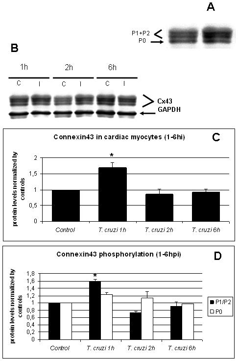
Figure 1. Initiation of infection induced a transient increase in Cx43 in cardiac myocytes.

A. Immunoblots for Cx43 of mouse cardiac myocytes had two upper phosphorylated bands (P1+P2) and one lower non-phosphorylated band (P0). B. Immunoblot analysis of non-infected (C) or T. cruzi-infected (Tc) cultured myocytes. GAPDH was used as loading control for all the experiments. C. Whole protein quantification of Cx43 showed that at one hour PI there was a 68% increase in Cx43 expression and at two and six hours, there was a 13.4% and 7.9% decrease respectively. D. Differential quantification for protein phosphorylation analyses of P1+P2 (phosphorylated) and P0 (non-phosphorylated) showed that both forms of Cx43 were transiently increased during the first hour of infection (60% and 20%, respectively). After this period, all three bands returned to levels near baseline (*: p<0.05, ANOVA).
