
Figure 2. T. cruzi alters cardiac myocyte Cx43 distribution at initial times of infection.
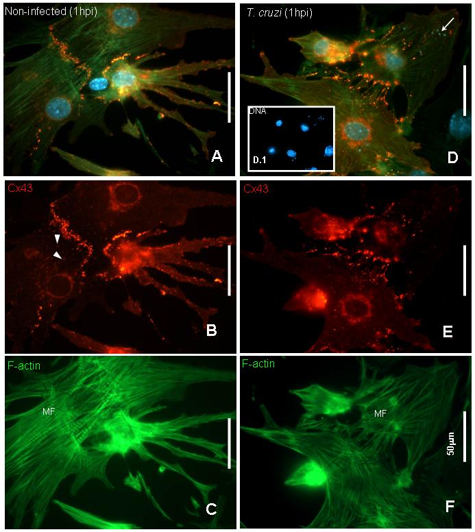
Mouse cardiac myocytes infected after 72 hours of cultivation were fixed at one hour post infection and stained with anti-connexin43 (Sigma antibody), Phalloidin-FITC for F-actin filaments and DAPI for DNA. A. Merged image of a non-infected culture showing that cardiac myocytes were connected by Cx43 and well differentiated. B. Cx43 was localized at cell-cell contacts and also displayed large vesicular structures (arrowheads). C. Phalloidin staining showed that cells were differentiated into myocytes with well defined polarity and presence of myofibrils (MF). D. and E: After one hour of infection Cx43 lost its plaque distribution compared with uninfected cells and was evident in stress fibers. In addition, there was more perinuclear staining indicating intense synthesis of Cx43. F. Phalloidin staining remained similar to observed in controls (C). D and D.1 show in detail DAPI staining corresponding to host cell and trypomastigotes’ DNA (arrows). Bars= 50.0μm.