
Figure 4. Immunostaining of mouse embryo cardiac myocyte gap junction protein Cx43.
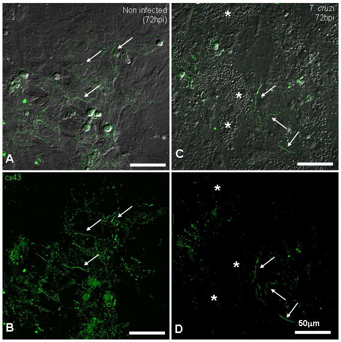
A and C show the merged image of DIC and anti-Cx43 antibody. Non-infected (A and B) cultures presented abundant gap junction plaques (arrows) between coupled cardiac myocytes. After 72 hours of infection (C and D), heavily infected cells (*) lost Cx43 staining and non-infected cells in the same field displayed strong junction plaques, similar to controls (arrows). Note in C, amastigote forms of the parasite revealed by DIC microscopy. (Scale bars = 50.0μm).