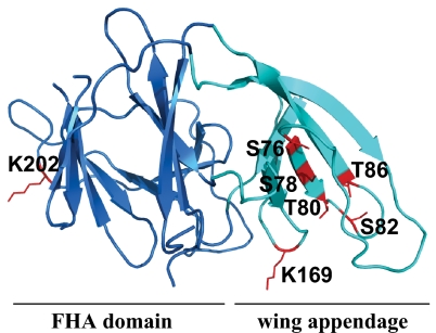
Fig. 8.
Location of the N-terminal phosphorylation sites and autoubiquitination sites in the structure of Pellino 1. A homology model of Pellino 1 was generated based on the recently published structure of Pellino 2 (30) using the program Modeller (37). Ser-76, Ser-78, Thr-80, Ser-82, and Thr-86 of Pellino 1 (shown in red) are located in a region of antiparallel β-sheet, termed the wing (turquoise), which is an appendage of the cryptic FHA domain (blue) thought to interact with phosphorylated IRAK1. Lys-169 and Lys-202 of Pellino 1, which were detected as sites of autoubiquitination, are also shown in red.

