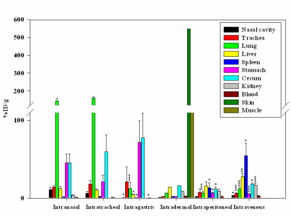
Figure 2.

Biodistribution of 64Cu labeled F. tularensis subsp. novicida. In order to confirm the observations from the microPET images the distribution of labeled bacteria was further analyzed by measuring the amount of radioactivity present in individual tissues at 20 hrs p.i. The data represent an average of 3–9 mice per route of infection and are expressed as % ID/g of tissue. The data obtained for each of the tissues sampled following i.n infection was compared statistically to the same tissue when inoculated by the i.g, i.p and i.v routes by using a two tail Student's t-test [e.g. lung (i.n) to lung (i.g)]. * = p < 0.05, ** = p < 0.01 and *** = p < 0.005. Data obtained following the i.d route was not included for the statistical analyses because it corresponded to just two biological replicates. The break in the scale was done between 110 and 120.
