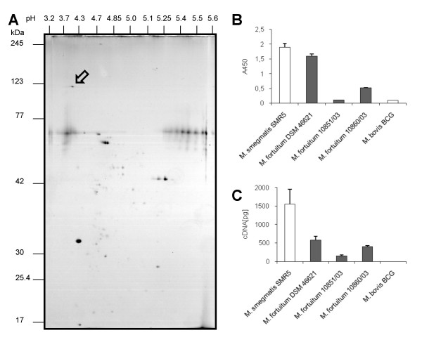
Figure 5.
Detection of PorMs in M. fortuitum and M. smegmatis. 2D-Electrophoresis, Western Blot, ELISA and qRT-PCR experiments proved PorMs to be expressed in the analysed strains. Section A shows 2D-Electrophoresis of protein isolation from the strain M. fortuitum 10860/03 using the detergent nOPOE. The arrow indicates the porin spot proven by Western Blot analysis (see Additional file 2). Section B and C show comparative analysis of porin expression among RGM. Expression of porin was detected by means of ELISA (B) and qRT-PCR (C). Each value represents the mean (± SD) of at least three independent experiments. B: Quantification of porin by means of ELISA in cell extracts of different mycobacteria using the polyclonal antibody pAk MspA#813. C: RT-Real-time-PCR quantification of porin mRNA in various RGM using specific primers and probes for mspA or porM, respectively.

