Figure 6.
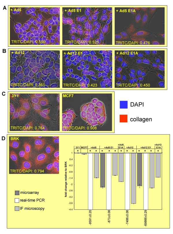
Expression of collagen type I observed using immunofluorescence microscopy. Immunofluorescence on cells stained for collagen type I (red) and nuclei staining with DAPI (blue) shows down-regulation of collagen as a result of transformation of BRK cells by Ad5 E1A (Panel A) or Ad12 E1A (Panel B) in comparison to untransformed rat fibroblast cell line 3Y1 and human breast tumour derived cell line MCF7 (Panel C). Representative images of one field are shown with corresponding TRITC:DAPI ratio for the individual field. Expression levels were calculated relative to intensity in untransformed BRK cells (Panel D). The dark grey bars represent the microarray results and the white bars represent the real-time PCR results while the light grey bars represent the immunofluorescence microscopy results. Each bar represents average ratio of intensity (corrected for background) of TRITC:DAPI for 10 fields imaged from same slide with significant results indicated by * (P < 0.500). Expression of collagen type I in MCF7 cells has been shown to vary according to the source of the cell line [86] and our results indicate variance on an individual cell basis (P 0.860).

