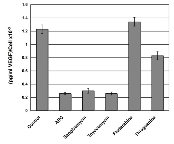
Figure 6.
ARC, sangivamycin and toyocamycin inhibit VEGF secretion. MCF7 cells were grown in 6 well dishes to 50% confluence, washed twice with PBS and 2 mL fresh RPMI-1640 media added. Media was then supplemented with the appropriate drug to a final concentration of 8 μM. After 24 h supernatant was removed and any cellular debris depleted by centrifugation. Adherent cells were trypsinized and cell numbers determined. An ELISA-based method was used to measure supernatant concentrations of VEGF. To avoid changes in cell number negatively influencing levels of VEGF secretion, results are expressed as picogram VEGF/mL per cell.

