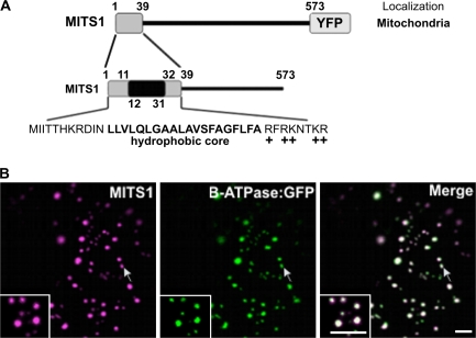
Fig. 1.
MITS1 harbours an N-terminal targeting signal and is localized to mitochondria. (A) Schematic representation of MITS1 and of its N-terminal region. Positively-charged residues follow a 20 residue hydrophobic region, characteristic of a mitochondrial targeting sequence. (B) In epidermal cells of tobacco leaves, MITS1:YFP labels punctate structures of various sizes that colocalize with the mitochondrial marker β-ATPase:GFP (arrows). Insets: magnified section of main panels. Scale bars=5 μm.

