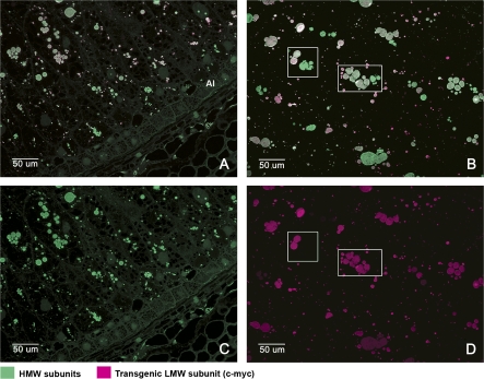
Fig. 4.
Immunofluorescence double labelling of the outer (A) and inner (B) endosperm of wheat grains at 20 dpa to show the locations of the tagged LMW subunit and HMW subunits. Alexa 568 (red) was conjugated to the anti-mouse secondary antibody recognizing the 9E10 antibody binding to the c-myc tag of the LMW subunit. Alexa 488 (green) was conjugated to an anti-rabbit secondary antibody recognizing the anti-R2-HMW antibody binding to HMW subunits. Micrographs (C) and (D) are single channel images corresponding to micrographs (A) and (B), respectively. The boxes in micrographs (B) and (D) show examples of protein bodies in the same cell which are labelled by the HMW antibody (green) but not by the anti-c-myc antibody (red). The fluorescence micrographs (A) and (C) correspond to the same region of the grain that is shown stained with toluidine blue in Fig. 1 D2. Fluorescence micrographs (B) and (D) correspond to those shown stained with toluidine blue in Fig. 1 D1.

