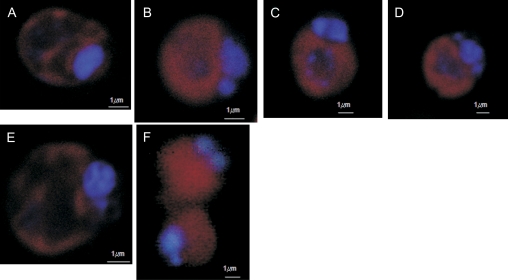
Fig. 3.
DNA condensation in Dunaliella viridis revealed by DAPI staining and confocal laser microscopy. (A) Control cells in PAR. (B) Cells after osmotic shock (5.5 M NaCl). (C) Cells after 4 h of UV radiation. (D) Cells after 2 h of heat shock. (E) Cells after 7 d of nitrogen starvation. (F) Culture senescence. Horizontal bar is 1 μm. (This figure is available in colour at JXB online.)

