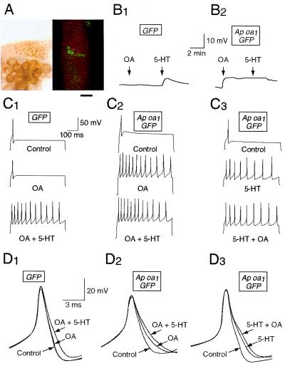
Figure 3.
Sample records of electrophysiological responses to OA and 5-HT in the sensory neurons expressing GFP (B1, C1, D1) or GFP plus Ap oa1 (B2, C2, C3, D2, D3). Resting potential (B1, B2), membrane excitability (C1–C3), and spike duration (D1–D3) were measured before (Control) and after drug applications. The cells B1, C1, and D1 were injected with pNEXδ-GFP, and the cells B2, C2, C3, D2, and D3 were injected with both pNEXδ-GFP and pNEXδ-Ap oa1. All these cells were GFP-positive. (A) Light (Left) and fluorescent (Right) photographs showing an example of GFP/Ap oa1-positive sensory neurons in a pleural ganglion. The cell bodies of these neurons are shown around the center of the fluorescent photograph. Bar = 250 μm. (B) Membrane depolarization by OA and 5-HT in a GFP-expressing cell (B1) and in a GFP/Ap oa1-expressing cell (B2). (C) Action potentials were recorded by injecting a depolarizing current (0.1–0.3 nA) for 500 ms. Membrane excitability was enhanced by 5-HT in a GFP-expressing cell (C1) and by OA and 5-HT in GFP/Ap oa1-expressing cells (C2, C3). (D) Superimposed action potentials demonstrating increases in spike duration after treatments with OA and 5-HT. Spike broadening by treatment with 5-HT in a GFP-expressing cell (D1) and with 5-HT and OA in GFP/Ap oa1-expressing cells (D2, D3). The shapes of single spikes were recorded by delivering a 0.4-nA current pulse for 15 ms. OA + 5-HT, 5-HT was applied later in the presence of OA (C1, C2, D1, D2). 5-HT + OA, OA was applied later in the presence of 5-HT (C3, D3).

