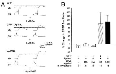
Figure 6.
Treatment with OA produced short-term facilitation in EPSP. (A) Representative monosynaptic EPSPs evoked by stimulating the sensory cells (SN) expressing GFP alone (Top) or expressing both GFP and Ap oa1 (Middle) or an uninjected sensory cell (SN) (Bottom). EPSPs were recorded at the motor neurons (MN) before (Left) and 5 min after (Right) the application of 1 μM OA (Top and Middle) or 10 μM 5-HT (Bottom) to the Aplysia pleural-pedal connections. (B) These group data indicate that OA enhanced amplitudes of EPSPs of motoneurons connected to sensory cells expressing Ap oa1. 5-HT also facilitated synaptic efficacy between uninjected sensory cell and motoneuron. Changes in EPSP amplitude are represented by blank bars (control for no activation of receptors), striped bar (activation of ectopic Ap oa1), and black bar (activation of endogenous 5-HT receptors). The height of each bar shows the mean ± SEM.

