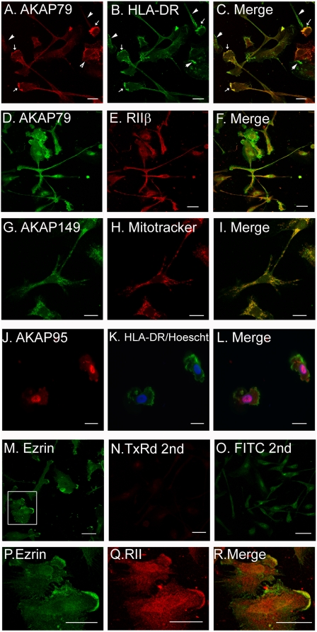
Figure 2. Immunofluorescence of AKAPs in dendritic cells.
Monocytes derived from adherence to plastic were plated onto poly-l-lysine coated glass coverslips. The cells were incubated for 30 minutes with mitotracker (panels H, I), and/or fixed, permeabilized, and stained with monoclonal antibodies to AKAP79 (panels A, C, D, F), AKAP149 (panels G, I), AKAP95 (panels J, L), HLA-DR (panels B, C, K, L), Ezrin (panels M, P), RIIβ (panels, E, F, Q, R). Hoescht (panel K, L) was added with secondary antibody conjugates. Panels P-R are 2 fold zoom of the boxed area of panel M. Scale bar = 25 µm.

