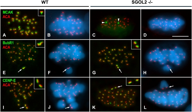
Figure 4. Distributions of MCAK, BubR1 and CENP-E in wild-type and Sgol2 −/− prometaphase I and metaphase I spermatocytes.
(A–D) Metaphase I spermatocytes double immunolabeled for MCAK (green) and kinetochores (ACA, red). MCAK is accurately located at the inner centromere domain below the closely associated sister kinetochores (inset) in wild-type (WT) spermatocytes, but delocalized as one or two cytoplasmic aggregates (arrowheads) in Sgol2 −/− spermatocytes. (E–H) Prometaphase I spermatocytes double immunolabeled for BubR1 (green) and kinetochores (ACA, red). In both wild-type and Sgol2 −/− spermatocytes, BubR1 preferentially labels the kinetochores (arrows) of unaligned bivalents. (I–L) Prometaphase I spermatocytes double immunolabeled for CENP-E (green) and kinetochores (ACA, red). CENP-E appears enriched at kinetochores (arrows) of unaligned bivalents. Both BubR1 and CENP-E appear at the outer kinetochore above the ACA signals (insets in E–L). All spermatocytes shown are projections of several focal planes, and are counterstained with DAPI (blue). Scale bar 10 µm.

