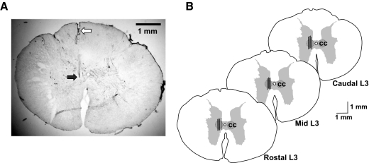
FIG. 6.
A: histological serial section of the rostral L3 spinal cord segment showing the tract (open arrow) and placement (filled arrow) of the tip of Eicom microdialysis probe. B: locations of microdialysis probes (black bars) implanted in the rostal, middle, and caudal regions of the Clarke's column nucleus (CC). Vertical lines represent the 1-mm length of dialysis membrane. The maximum field potential evoked by stimulation of the anterior cerebellar lobule corresponding to the location of Clarke's column was used as a guide for stereotaxic placement of the dialysis probes (see also Fig. 1A).

