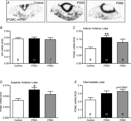
Figure 3. Pituitary pro-opiomelanocortin (POMC) mRNA expression in male offspring whose mothers were exposed to stress at PS50 and PS60 (as described in Fig. 1), or left undisturbed throughout pregnancy (control).
A, representative in situ hybridization images of POMC mRNA expression in control, PS50 and PS60 pituitary. Relative optical density (ROD) of glucocorticoid receptor (GR) mRNA in the anterior lobe (B), POMC mRNA in the inferior anterior lobe (C), POMC mRNA in the superior anterior lobe (D) and POMC mRNA in the intermediate lobe (E). Animal numbers are indicated within bars. *P < 0.05, **P < 0.01, compared to control. IL, intermediate lobe; AL, anterior lobe; Sup, superior anterior lobe; Inf, inferior anterior lobe.

