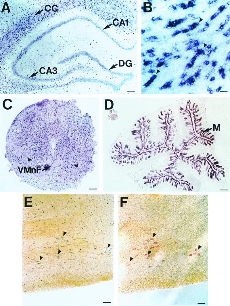
Figure 4.

Distribution of rCTL1 in adult rat tissues by ISH. (A) Intense labeling with the antisense rCTL1 cRNA probe was observed within scattered cells present at a higher density in the corpus callosum (CC) than in cells located in the gray matter, as well as in the hippocampal neurons of the Ammon's horn (CA1–CA3) and of the dentate gyrus (DG). (B) A higher magnification of labeled cells within the CC showed chains of cells suggesting the labeling of oligodendrocytes (arrowheads). (C) A frontal section of cervical spinal cord showed a high density of small labeled cells in both white and gray matter, and larger labeled cells in the ventral horns (arrowheads); VMnF, ventral median fissure. (D) The cell layer constituting the colonic mucosa (M) expressed high levels of rCTL1 transcript. (E and F) Double ISH using the digoxigenin-labeled rCTL1 cRNA antisense probe (E) and a fluorescein-labeled choline acetyltransferase antisense riboprobe (F) identify the large cells expressing rCTL1 as motor neurons (arrowheads). Cryosections hybridized with digoxigenin-labeled and fluorescein-labeled sense probes showed no significant signal. Bars correspond to 200 μm (A, C, and D), 25 μm (B), and 100 μm (E and F).
