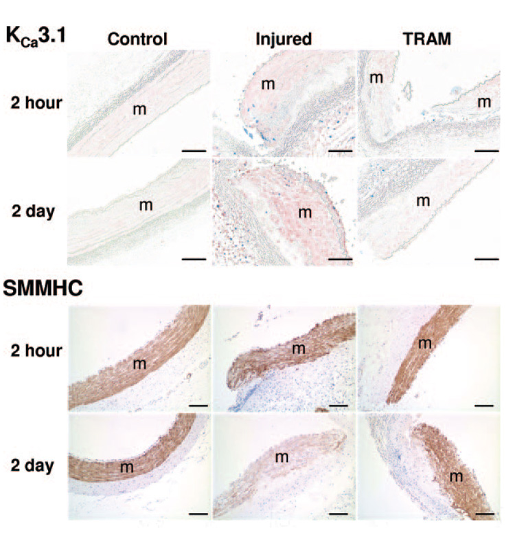
Figure 3. KCa3.1 and SMMHC histology 2 hours and 2 days postangioplasty.
Representative cross-sections (8 µm; 4 to 5 per group) of control, injured, and TRAM-34–coated balloon injured (TRAM) LCX and LAD 2 hours and 2 days postangioplasty exposed to antibodies against KCa3.1 (1:600, pink, top), Ki-67 (1:200, blue, top), and SMMHC (1:800, brown, bottom). Horizontal bar=100 µm.

