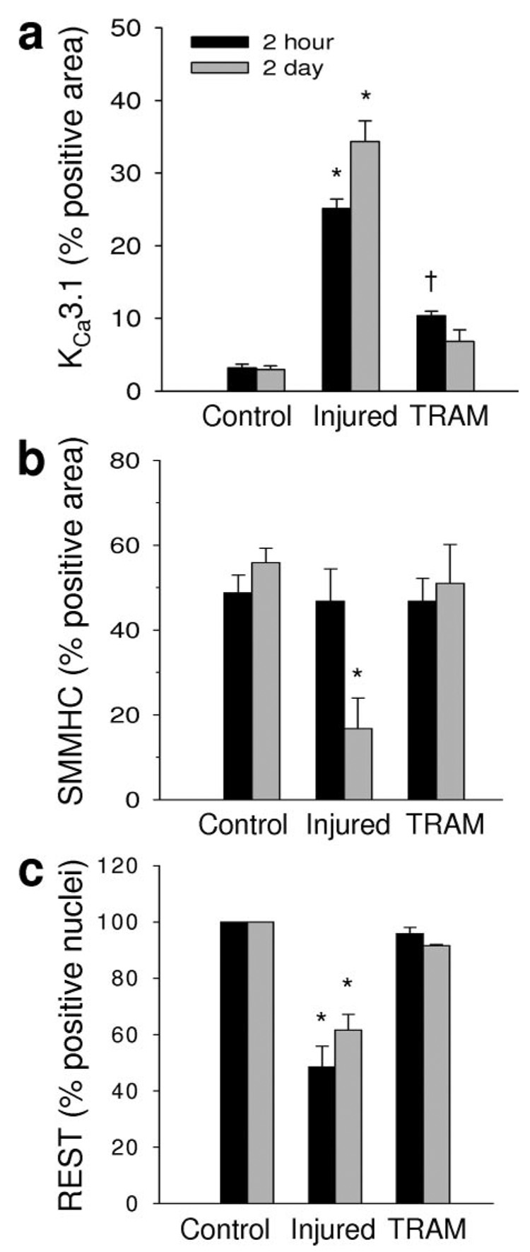
Figure 5. Histological analysis of KCa3.1, SMMHC, and REST.
KCa3.1 (a), SMMHC (b), and REST (c) staining were quantified using Image Pro Plus. Injury-induced changes in KCa3.1, SMMHC, and REST staining were blocked by TRAM-34 (see Figure 3 and Figure 4 for representative images). *P<0.05 vs respective control and †P<0.05 vs injured and control (n=4 to 5 per group).

