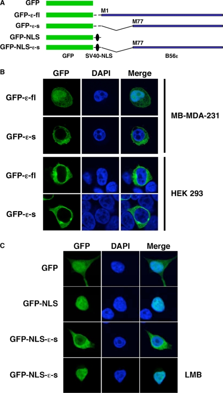
FIGURE 7.
Subcellular localization of B56ε isoforms. A, schematic drawing of constructs used in B and C. B, N-terminal GFP-tagged B56ε-fl and N-terminal GFP tagged B56ε-s were transfected into MB-MDA-231 breast cancer and HEK 293 cells. Transfected cells were stained with DAPI and subjected to confocal microscopy analysis. Columns from left to right are GFP-tagged proteins, DAPI staining, and merged images. GFP-B56ε-s is restricted to the cytoplasm in both cell lines. In MB-MDA-231, GFP-B56ε-fl was detected in both cytoplasm and nucleus. In HEK 293 cells, it is mainly localized in the cytoplasm. C, subcellular localization of GFP, GFP-NLS, and GFP-NLS-ε-s in HEK 293 cells. The bottom panels are GFP-NLS-ε-s in cells treated with leptomycin B (LMB). Columns from left to right are the distribution of GFP or GFP-tagged proteins, DAPI staining, and merged images. Note that GFP-NLS was nucleus-localized, whereas GFP-NLS-ε-s was detected in both nucleus and cytoplasm. When treated with leptomycin B, GFP-NLS-ε-s became nucleus-localized.

