FIGURE 3.
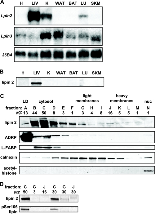
Lipin 2 is a hepatic-enriched lipin protein. A, the autoradiographs depict representative northern blots showing expression of Lpin2, Lpin3, or 36B4 (to control for loading) in WT mouse tissues (heart (H), liver (LIV), kidney (K), white adipose tissue (WAT), brown adipose tissue (BAT), lung (LU), skeletal muscle (SKM)). B, representative Western blots performed using a polyclonal antibody against lipin 2 and tissue lysates from WT mice are shown. C, the images depict results of Western blotting studies using hepatic lysates from WT mice fractionated by gradient centrifugation and probed with antibodies listed at left. LD, lipid droplet. The fractions are lettered alphabetically from the top to the bottom of the gradient. The gel was loaded by equal volumes, so the quantity of protein in each lane varies as is listed above each lane. L-FABP, liver fatty acid binding protein; nuc, nuclear. D, fractions C, G, and J from panel C were used in these Western blot studies. Each lane was loaded by volume (lanes 1–3) or by equal protein (lanes 4–6; 30 μg). Blots were then probed with antibodies against lipin 2 or phosphoserine 106 lipin.

