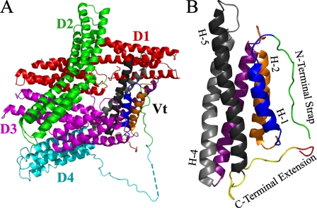
FIGURE 1.
A, a ribbon diagram illustrating the structure of full-length vinculin (PDB ID 1ST6). In the closed, autoinhibited conformation, the clamp-like head domain (Vh, D1 (red), D2 (green), D3 (magenta), and D4 (cyan)) forms a tight interaction with the tail domain (Vt, (multi-color)). Current models of vinculin activation and function require the release of the head/tail interaction to allow ligand binding. B, a ribbon diagram illustrating the isolated tail domain of vinculin (PDB ID 1ST6). The N-terminal strap and C-terminal extension are highlighted (green and yellow, respectively). The hydrophobic hairpin at the extreme C terminus is shown in red. Select helices are labeled (e.g. H-1), and for clarity, the helices are colored identically in parts A and B. A more detailed illustration, highlighting specific interactions between the N-terminal strap and C-terminal extension, is shown in Fig. 7.

