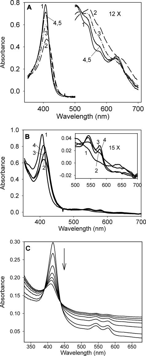
FIGURE 5.
Reaction of KatG[W107F] with peroxides. A, spectral changes upon addition of 30 μm PAA to 10 μm KatG[W107F] in 20 mm phosphate buffer, pH 7.2. 1, resting enzyme; 2, t = 1 s; 3, t = 22 s; 4, t = 100 s; 5, t = 200 s. B, spectral changes of 10 μm KatG[W107F] upon addition of 50 μm H2O2 in 20 mm phosphate buffer, pH 7.2, at 25 °C. 1, resting enzyme; 2, t = 0.5 s; 3, t = 5 s; 4, t = 105 s. C, heme breakdown during long term incubation of KatG[W107F] with excess H2O2. M. tuberculosis KatG[W107F] (4 μm) was mixed with 1 mm H2O2 in 20 mm phosphate buffer, pH 7.2, at 25 °C. Spectra were recorded at 1, 6, 12, 24, 36, 48, 60, and 84 min after mixing.

