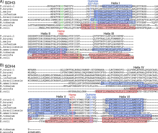
FIGURE 5.
Alignments of SDH3 (A) and SDH4 (B) sequences. Amino acid residues proposed for binding of protoheme IX are shown in red and those for the quinone binding in blue. Other conserved residues are indicated by green. Transmembrane helices found in E. coli (Protein Data Bank code 1NEK) and porcine (Protein Data Bank code 1ZOY) Complex II are shown by red rectangles, and transmembrane helices predicted by TMHMM are indicated by blue rectangles. TMHMM failed to predict transmembrane helices in T. brucei SDH3. Residue numbers refer to E. coli SDH3 (SdhC) and SDH4 (SdhD). GenBank™ accession numbers for SDH3 and SDH4 sequences used are T. cruzi (XP_809410, XP_808211), T. brucei (XP_845531, XP_823384), L. major (XP_001684890, XP_001685874), L. infantum (XP_001467132), L. brasiliensis (XP_001566908, XP_001567905), R. americana (NP_044796, NP_044797), Nicotiana tobacum (YP_173376, YP_173457), Sus scrofa (1ZOY_C, 1ZOY_D), and E. coli (NP_415249, NP_415250).

