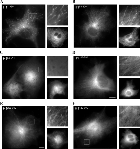
FIGURE 2.
Plus-end tracking behavior of CLIP-170 fragments in vivo. COS-7 cells were transfected with the GFP-tagged plasmid constructs described in Fig. 1A. Single frames from movies have been extracted for presentation here. Right upper panels have been magnified 2.5× from each boxed region. Right lower panels represent the same construct at a high level of expression as measured by fluorescence intensity. A, full-length head domain H1-(1–350) tracks microtubule plus ends. B, H1-(58–300) tracks plus ends in most transfected cells and decorates the MTs in overexpressing cells to similar levels as H1-(1–350). C, H1-(58–211) does not track microtubule plus ends and remains diffuse throughout the cytoplasm at all levels of expression. D, H1-(156–350) has some ability to localize to the MT lattice but no real preference for the plus end. E, H1-(203–350) tracks microtubule plus ends, although with less robust tips compared with either construct containing two CAP-Gly domains; this construct does bind the lattice at higher levels of expression. F, H1-(122–300) is largely diffuse, localizing only weakly to MT fibers, and was not observed to track MT plus ends. (Scale bar, 10 μm.)

