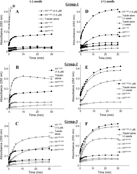
FIGURE 3.
Assembly promoting activity of CLIP-170 fragments. A–C, MT assembly in the presence of CLIP-170 fragments was monitored by change in the absorbance at 350 nm. Polymerization of tubulin (12 μm) was measured in the absence and presence of the different CLIP-170 fragments (2.0 μm) as shown. In this assay, H1-(1–350) (2.0 μm) causes strong bundling and thus produces an off-scale light scattering signal (A, asterisk). To minimize this effect and for comparisons with the other CLIP-170 fragments, H1-(1–350) was also used at 1.0 μm as shown. Data were extracted at different time points and plotted using the GraFit 6 program; the lines are provided as a guide. Each data point is the average from two independent experiments. D–F, polymerization with or without CLIP-170 fragments as shown was carried out in the presence of taxol seeds (1.0 μm) to observe the effects on microtubule elongation. The tubulin concentration was 12 μm and all CLIP-170 fragments were used at 1.0 μm concentration to minimize possible bundling effects.

