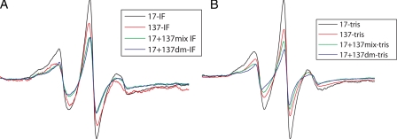
FIGURE 2.
Comparison of EPR spectra for single mutants (17-17 and 137-137), 17 + 137 mixture, and 17 + 137 double mutant showing the mobility of the spin labels and dipolar interaction. The double mutant (17 + 137 dm) showed greater broadening of spectra emphasizing the strong interaction at these positions in head and rod domain. A, EPR spectra scanned when spin-labeled proteins were dialyzed against IF assembly buffer and against low ionic strength Tris buffer (B).

