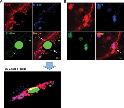
FIGURE 4.
Co-localization of MTS15, FSP1, aP2, and LipidTox in thymus of FoxN1Cre;ROSA26RstoplacZ mice. A, thymic cryosections of 6-month-old FoxN1Cre; ROSA26RstoplacZ labeled with MTS15 (fibroblast marker), β-galactosidase (red), and LipidTox (green). The confocal Z-stack image reconstruction in three-dimensional (3D) orientation depicts the presence of LipidTox-stained cells within the thymic subcapsule. The merge ofβ-galactosidase (red) and MTS15 (blue) appears as cyan cells enveloping the large unilocular lipid vacuole. B, the subcapsular region of thymus showing co-localization of β-galactosidase (red), FSP1 (blue), and aP2 (green).

